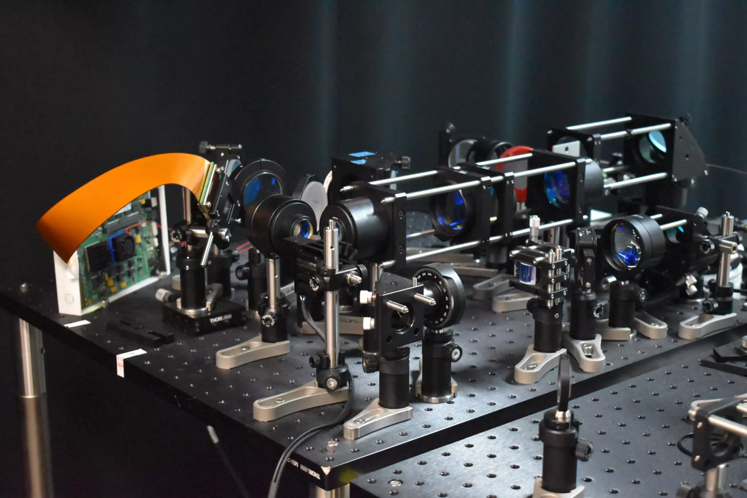
The advancement of science and technology continues to push the boundaries of what we know about the human brain. Recently, researchers developed a cutting-edge two-photon fluorescence microscope that has the capability to capture high-speed images of neural activity at the cellular level. This innovative approach is set to revolutionize the field of neuroscience by providing a clearer view of how neurons communicate in real time, thus shedding light on brain function and neurological diseases.
Traditionally, two-photon microscopy has been used to image deep into brain tissue by scanning a small point of light across the sample area to excite fluorescence and collect signals point by point. While this method provides detailed images, it is slow and can potentially harm brain tissue. The new microscope takes a different approach by incorporating a new adaptive sampling scheme and replacing point illumination with line illumination. This enables in vivo imaging of neuronal activity in a mouse cortex at speeds ten times faster than traditional two-photon microscopy, while reducing the laser power on the brain more than tenfold.
The key innovation behind the new microscope is the adaptive sampling strategy that uses a short line of light to illuminate specific regions of the brain where neurons are active. By targeting active neurons without capturing background or inactive areas, the total light energy deposited on the brain tissue is significantly reduced, minimizing the risk of damage. This dynamic approach is made possible by utilizing a digital micromirror device (DMD) to shape and steer the light beam with precision, allowing for fast and accurate targeting of active neuronal regions.
One of the major achievements of the new microscope is the ability to capture calcium signals, which indicate neural activity, in living mouse brain tissue at a speed of 198 Hz. This is a significant improvement over traditional two-photon microscopes and enables the monitoring of rapid neuronal events that may be missed by slower imaging methods. The adaptive line-excitation technique, combined with advanced computational algorithms, allows for the isolation of individual neuron activity, providing a deeper understanding of complex neural interactions and the functional architecture of the brain.
Moving forward, researchers are focused on integrating voltage imaging capabilities into the microscope to provide a direct and rapid readout of neural activity. This will open up new possibilities for studying brain function in real time and exploring neural activity during learning and disease states. Additionally, efforts are being made to improve the user-friendliness of the microscope and reduce its size to make it more accessible for neuroscience research.
The development of the high-speed two-photon fluorescence microscope marks a significant milestone in the field of neuroscience. By offering a powerful imaging tool that enables the study of dynamic neural processes in real time with minimal risk to living tissue, researchers are poised to uncover new insights into brain function and neurological diseases. The future of neuroscience is bright, thanks to the technological innovations that continue to push the boundaries of our understanding of the brain.


Leave a Reply
You must be logged in to post a comment.