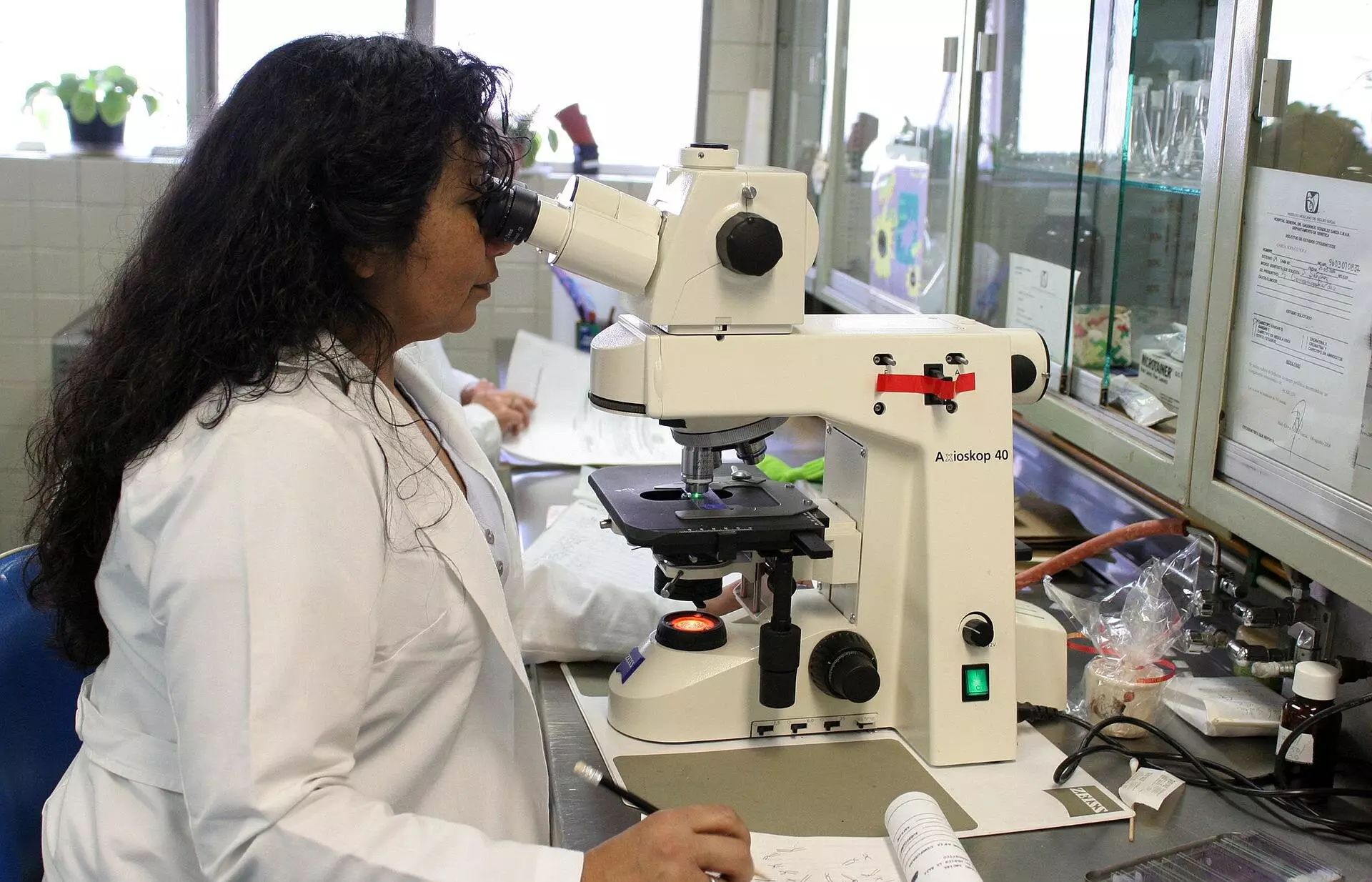
When using a microscope to view biological samples, it is essential to consider the medium in which the lens of the objective is situated. The disturbance in the light beam occurs when the lens is in a different medium than the sample being observed. This discrepancy in mediums causes the light rays to bend differently, leading to inaccuracies in the measurement of the depth of the sample. Consequently, the sample appears flattened as a result of this optical distortion.
An Evolution in Corrective Factor Theories
Traditionally, theories developed in the 80s proposed a constant corrective factor to determine the depth of the sample, regardless of its actual depth. However, Nobel laureate Stefan Hell pointed out in the 90s that this scaling factor may vary depending on the depth of the sample. Sergey Loginov, a former postdoc at Delft University of Technology, has recently demonstrated through calculations and a mathematical model that the sample appears more flattened closer to the lens than further away. This discovery challenges the longstanding assumption of a constant corrective factor and highlights the importance of considering depth-dependent scaling in microscopy.
Following Loginov’s work, Ph.D. candidate Daan Boltje and postdoc Ernest van der Wee conducted experiments in the lab to validate the depth-dependent corrective factor. Their findings, published in the journal Optica, confirmed the significant impact of depth on the appearance of the sample under a microscope. Van der Wee emphasizes the practical implications of their research by providing a web tool and software that allow researchers to determine the precise corrective factor for their specific experiments.
“With the aid of our calculation tools, we can now accurately extract a protein and its surroundings from a biological system for analysis with electron microscopy,” explains Boltje. This level of precision is crucial in microscopy, where mistakes can be costly and time-consuming. By optimizing depth determination, researchers can streamline their experiments, reduce costs, and focus on studying relevant proteins and biological structures. Understanding the precise structure of proteins in biological systems is key to advancing our knowledge of diseases and abnormalities, paving the way for potential therapeutic interventions.
The web tool developed by the research team allows users to input specific details of their experiment, including refractive indices, aperture angle of the objective, and wavelength of the light used. The tool then generates a curve illustrating the depth-dependent scaling factor, enabling researchers to account for optical distortions accurately. This data can be exported for further analysis and comparison with existing theories, providing valuable insights into the behavior of light in microscopy.
The recognition of depth-dependent scaling factor in microscopy represents a significant advancement in the field of biological imaging. By acknowledging and accounting for the optical distortions caused by varying depths, researchers can improve the accuracy and efficiency of their experiments. The development of tools like the web tool presented in this study highlights the value of interdisciplinary collaboration in tackling complex scientific challenges and advancing our understanding of the microscopic world.


Leave a Reply
You must be logged in to post a comment.