
Nonlinear light microscopy has transformed our ability to observe and comprehend complex biological processes. This innovative technology has opened new doors in the field of biology, allowing for detailed insights into cellular functions and interactions. However, despite its numerous benefits, light microscopy also poses a potential risk – the damaging effects of intense light on living matter.
Researchers at the Max Planck Institute for the Science of Light and Max-Planck-Zentrum für Physik und Medizin have come together to investigate the mechanisms behind the irreversible perturbation of cellular processes caused by intense light exposure. This collaborative effort aims to identify the conditions under which intense pulsed lasers can be safely used in vivo without causing harm to the organism.
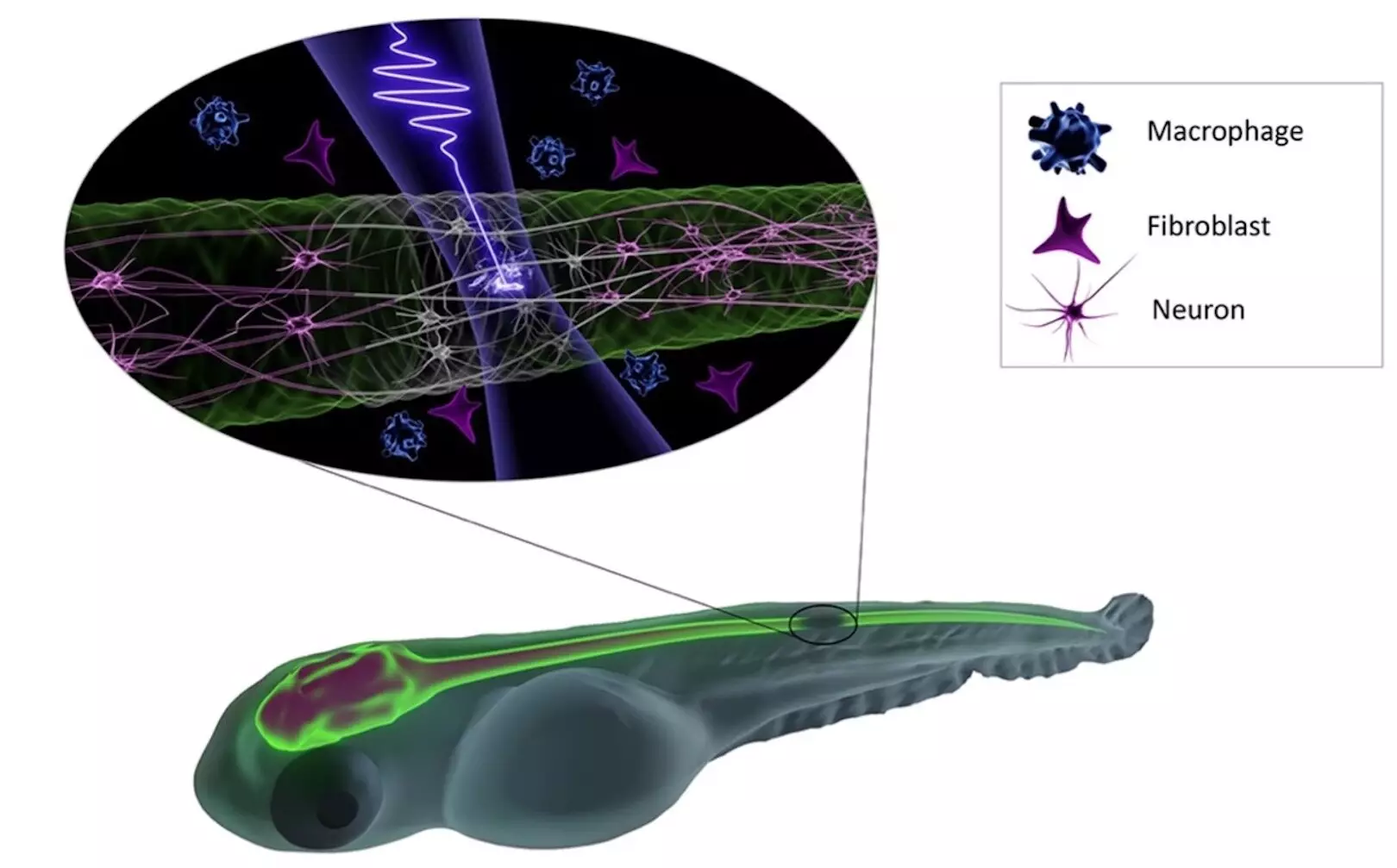
Using the vertebrate species zebrafish as a model organism, the international research team explored the effects of photodamage in deep tissue at a cellular level induced by femtosecond excitation pulses. The study revealed that damage to the zebrafish central nervous system occurs abruptly at extreme peak intensities required for low-density plasma formation.
The findings of this research have significant implications for the field of nonlinear label-free microscopy. By understanding the thresholds for photodamage, researchers can now increase the imaging dwell time and photon flux during irradiation at specific wavelengths while avoiding detrimental effects on living tissues.
The development of innovative microscopy techniques, such as femtosecond fieldoscopy, offers exciting opportunities for high-resolution, label-free imaging with attosecond temporal resolution. These advancements not only enhance our understanding of biological processes but also pave the way for precise manipulations of the central nervous system using light-based technologies.
The collaborative nature of this research project highlights the importance of interdisciplinary collaborations between the fields of physics and biology. By combining expertise from different disciplines, researchers can make significant strides in advancing our understanding of complex biological phenomena and developing cutting-edge imaging technologies.
The study of photodamage in deep tissue imaging has provided valuable insights into the limitations and possibilities of intense light exposure in biological systems. By unraveling the mechanisms behind photodamage, researchers can optimize imaging techniques and explore new avenues for noninvasive manipulation of cellular processes. This research not only contributes to the field of microscopy but also underscores the power of interdisciplinary collaborations in driving scientific innovation and discovery.
Leave a Reply
You must be logged in to post a comment.